Birth Defects
Causes of defects, Physical birth defects, Hereditary diseases and syndromes
Birth defects or congenital defects are present at birth. They result from heredity, environmental influences, or maternal illness. Such defects range from the very minor, such as a dark spot or birthmark that may appear anywhere on the body, to more serious conditions that may result in marked disfigurement, impaired functioning, or decreased lifespan.
A number of factors individually or in combination may cause birth defects. Heredity plays a major role in passing birth defects from one generation to the next. Inherited conditions are passed on when a baby receives a flawed gene from one or both parents. Conditions such as sickle cell anemia, color blindness, deafness, and extra digits on the hands or feet are hereditary. The condition may not appear in every generation, but the defective gene usually is passed on. A classification of structural defect can be as follows: Malformation (poor formation), deformation (due to fetal constraint that can result in damage (e.g., central nervous system damage or limb reduction) and disruption of previous normally formed structures (due to vascular damage, vascular exchange of necrotic debris).
Low birth weight deriving from a fetal growth restriction (FGR) is the most common birth defect, with one in every 15 babies being born at less than their ideal weight. A baby whose weight lies in lowest 10% of the normal population is designated as having a FGR. At term of pregnancy, a baby who weighs 5 lb, 8 oz (2,500 g) at birth has a low birth weight. One who is born weighing 3 lb, 5 oz (1,500 g) has a very low birth weight. A low birth weight baby born after a normal gestation period is called a small-for-date or small-for-gestational-age baby.
Exposure of the mother to chemicals such as mercury or to radiation during the first three months of pregnancy may result in an abnormal alteration in the growth or development of the fetus. The mother's diet may also be a factor in her baby's birth defect. A balanced and healthy diet is essential to the proper formation of the fetus because the developing baby receives all of its nutrition from the mother.
Prenatal development of the fetus may also be affected by disease that the mother contracts, especially those that occur during the first trimester (three months) of pregnancy. For example, if a pregnant woman catches rubella, the virus crosses the placenta and infects the fetus. In the fetus, the virus interferes with normal metabolism and cell movement and can cause blindness (from cataracts), deafness, heart malformations, and mental retardation. The risk of the fetal damage resulting from maternal rubella infection is greatest during the first month of pregnancy (50%) and declines with each succeeding month.
It is especially important that the mother not smoke, consume alcohol, or take drugs while she is pregnant. Drinking alcohol heavily can result in fetal alcohol syndrome (FAS), a condition that is physically apparent. FAS newborns have small eyes and a short, upturned nose that is broad across the bridge, making the eyes appear farther apart than normal. These babies also are underweight at birth and do not catch up as time passes. They often have some degree of mental retardation and may exhibit behavior problems. A mother who continues to take illicit drugs such as heroin, crack, or cocaine will have a baby who is already addicted. The addiction may not be fatal, but the newborn may undergo severe withdrawal, unless the addiction is revealed and carefully treated. Furthermore, some behavior problems/cognitive deficits are suspected to be associated with fetal drug exposure and addiction.
Some therapeutic drugs taken by pregnant women have also been shown to produce birth defects. The most notorious example is thalidomide, a mild sedative-hypnotic agent. During the 1950s women in more than 20 countries who had taken this drug gave birth to more than 7,000 severely deformed babies. The pattern of malformation seen in affected infants included phocomelia, polydactyly, syndactyly, facial capillar hemangiomas, hydrocephalus, renal anomalies, cadiovascular anomalies, ear and eye defects, and intestinal anomalies. The principal defect these children suffered is phocomelia, characterized by extremely short limbs often with no fingers or toes.
Clubfoot
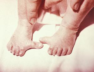
Approximately one newborn out of every 735 has a form of clubfoot. In the most serious form, known as equinovarus, the foot is twisted inward and downward and the foot itself is cupped or flexed. If both feet are clubbed in this manner the toes point to each other rather than straight ahead. Often the heel cord or Achilles tendon is taut so that the foot cannot be straightened without surgery.
A milder and more common type of clubfoot is called calcaneal valgus, in which the foot is bent upward and outward in the same way that you would flex your foot at the ankle. Still other forms include the talipes cavus in which the instep is abnormally elevated; talipes valgus in which the heel is turned outward, and talipes varus in which the heel is turned inward.
The seriously deformed clubfoot requires surgery to realign the bones and ligaments. The milder forms often can be cured by fitting the baby with corrective shoes to gradually move the bones back into alignment.
Cleft lip and cleft palate
Approximately 7,000 newborns (one of every 930 births) are born with cleft lip and/or cleft palate each year in the United States. Cleft lip and palate describe a condition in which a split remains in the lip and roof of the mouth. Although cleft lip and palate are two distinct anomalies, they frequently occur together. Cleft lip with or without cleft palate occurs in 60-75% of the cases. twenty-five to forty percent are isolated cleft palate. During growth in utero (in the womb) the lip or palate, which develop from the edges toward the middle, fail to grow together. Such a failure is a consequence of the abnormal migration and proliferation of facial embryonic tissues called mesenchyme. The defect occurs most often among Asians and certain Native American groups, less frequently among whites, and least often among African Americans.
Approximately 25% of infants born with cleft palate have inherited the trait from one or both parents. The cause for the other 75% remains unknown, but may be a combination of heredity, poor nutrition, use of drugs, or a disease the mother contracted while pregnant. Maternal smoking represents the most controversial association. The cleft may involve only the upper lip, may extend into the palate, or may be located on the back of the palate.
Surgery is especially important to correct the defect in the palate. Feeding a baby with cleft palate is difficult because the food can pass through the palate into the nasal cavity and may be inhaled and cause choking. In the newborn, whose bones have not completely hardened, surgery is relatively simple. As the child ages, however, surgical correction is more difficult and the child will require speech therapy.
Spina bifida
Spina bifida or open spine occurs once in 2,000 births in the United States. It belongs to a group of defects known as neural tube defects that are the second most prevalent neonatal anomaly in the United States after cardiac malformations. It occurs when the edges of the spine that should grow around the spinal cord do not meet. An open area remains, which can mean that an area of the spinal cord (or the entire spinal cord, in the most severe cases) are unprotected. The mildest form of spina bifida may be so slight that the defect does not have any effect on the child and is discovered by accident, usually when an x ray is taken for another reason. The term spinal bifida means the spine is cleft, having an opening or space, in two parts.
Spina bifida may present itself as a cyst, ranging in size from a walnut to a grapefruit, in which some parts of the meninges (layers of connective tissue covering the spinal cord), spinal cord, or both are contained. The lump can be removed surgically. In the most serious form, the lump or cyst has little skin or covering so spinal fluid may leak from it. Roots of the spinal nerves are contained within the cyst and the cyst may be covered with sores. Infection is a serious risk until surgery has been performed and the area has healed. Unfortunately, this condition may leave the child's legs partially or completely paralyzed and without feeling. Other associated problems may include control of the bowels and bladder.
Newborns with spina bifida often have an associated condition called hydrocephalus, which literally means water in the head. In this condition, cerebrospinal fluid collects in and around the brain and will not drain. Mental retardation can result if the fluid is not drained regularly. This can be accomplished by implanting a special tube (called a shunt) leading from the brain down into a vein in the child's neck or into the child's chest to allow the fluid to drain harmlessly. Hydrocephaly also can occur in infants who do not have spina bifida. The cause of spina bifida is not known, nor is any means of prevention. It can be diagnosed before birth by amniocentesis (by dosing the intra-amniotic alpha feto protein) or ultrasound. The risk of having a baby with spina bifida or other associated defects seems to be reduced if a woman takes at least 400 mg of folic acid just before and throughout pregnancy.
Heart defects
Congenital heart defects occur in one of every 115 births in the United States. The defect may be so mild that it is not detected for some years or it may be fatal. A baby with a heart defect may be born showing a bluish tinge around the lips and on the fingers. This condition, called cyanosis, is a signal that the body is not receiving enough oxygen. The blue color may disappear shortly after birth, indicating that all is normal, or it may persist, indicating that further testing is needed to determine the nature of the heart defect.
A normal heart has four chambers; two upper, called the atria (singular: atrium) and two lower called the ventricles. The right heart receives the blood that is returning from the body, and has been depleted of oxygen. This oxygen-poor blood arrives in the right atrium, where it is pumped into the right ventricle. The right ventricle sends oxygen-poor blood to the lungs, where it is exposed to and picks up plenty of oxygen again. This oxygen-rich blood enters the left atrium and is then pumped into the left ventricle. The left ventricle pumps oxygen-rich blood through the aorta to all the organs and tissues of the body.
During fetal development, blood circulation occurs differently, because the fetus' blood does not need to flow through its lungs. It receives its oxygen from the mother through the placenta via the umbilical cord. Since the atria communicate during fetal life, blood rich in oxygen coming from inferior vena cava crosses the foramen ovale and into the left atrium bypassing the lungs (eventually the foramen ovale is closed from the higher pressure generated at the left side after the lunds expand at birth). Another special shunt, the arterious duct connects the main pulmonary artery to the aorta. In such a way, the blood flow that does enter the right atrium enters the right ventricle, then the main pulmonary artery, then enters the ductus arteriosus which connects to the aorta. In this way the vast majority of blood flow bypasses the lungs during development of the fetus. Normally the shunts should close at birth. After birth, blood should begin to circulate through the lungs for the first time, because the newborn baby's lungs are now responsible for delivering oxygen to the blood. Sometimes, however, the shunt does not close properly, and blood is not appropriately circulated through the lungs. When this occurs, surgery is required to close the shunt and restore normal circulation.
If it is undetected at birth, a heart defect may impair the growth of a child. He will be unable to exert the energy that other children do at play because he cannot supply sufficient oxygen to his body. He may become breathless at small amounts of exertion and may squat frequently because it is easier to breathe in that position.
Some minor defects may disappear over time as the child grows. A small hole in the wall between the left and right sides of the heart, which causes symptoms by allowing the mixing of oxygen-poor and oxygen-rich blood, for example, may spontaneously close over time. A larger defect will require surgical patching.
Some newborns may have only one upper chamber or only a single lower chamber of the heart. The aorta, where it begins at the heart, may be narrowed (stenosed) and impair the flow of blood from the heart. Some of the heart valves may not function correctly and occasionally the vessels of the heart may be transposed so that the aorta leads from the right side of the heart, delivering oxygen-poor blood to the organs and tissues.
These are only a few of the heart anomalies that can be present in the newborn. The heart is a complicated organ and its formation can be influenced by hereditary factors as well as by alcohol consumption or smoking. Fortunately, most heart defects correct themselves over time or can be corrected with surgery.
Other physical deformities
Physical defects in newborns are common. They can affect any of the bones or muscles in the body and may or may not be correctable. Among the more common are the presence of extra fingers or toes (polydactyly), which presents no health threat and can be corrected surgically. Similarly, webbed fingers and toes, a genetic disorder, seen in approximately one of every 1,700 to 2,000 births, can be treated surgically to resemble a normal appendage.
A more serious, though relatively rare, condition is called achondroplasia; this term means without cartilage formation and refers to the supposed lack of cartilage growth plates near the ends of a child's bones. In fact, the plates are present, but grow poorly. Achondroplasia is a type of dwarfism. This genetic disorder of bone growth is seen in one in 20,000 births and is one of the oldest known birth defects. Ancient Egyptian art shows individuals with this condition.
The cause of achondroplasia is not known, nor is there a cure. The child who has this condition will be slow at walking and sitting because of his short arms and legs, and this may be interpreted as mental retardation. However, these individuals have normal intelligence.
In addition to physical deformities, certain diseases and syndromes also are passed to the infant through the parents' genes. Some of these conditions can be controlled or treated while others are untreatable and fatal.
Sickle cell anemia
Sickle cell anemia is an inherited disease of the blood cells that occurs in about one of every 400 African Americans. An individual can be a carrier of sickle cell anemia, in which case he or she has the gene but does not show any active signs of the disease. If two carriers become parents, however, some of their children may have sickle cell anemia.
The disease gets its name because certain red blood cells assume a sickle shape and lodge in small blood vessels. This altered shape is a function of the hemoglobin molecule present in red blood cells. Two forms of hemoglobin make up these cells: hemoglobin A (Hb A) and hemoglobin B (Hb B). In individuals with sickle cell anemia, Hb B is instead produced as Hb S, a form of hemoglobin with a rigid, sickle shape that deforms the red blood cell. When the cell becomes wedged in a small blood vessel it prevents the flow of blood through the vessel and can initiate what is called a sickle cell crisis. The lack of blood flow to the tissues being blocked causes pain and inflammation of the oxygen-deprived tissue.
Abnormal red blood cells are removed from the circulatory system by the spleen, but removal of large numbers of such cells can lead to anemia, a lack of adequate numbers red blood cells. Unfortunately, the breakdown of abnormal red blood cells can in itself cause a serious condition in which excess iron, scavenged from the hemoglobin molecule, is deposited in tissues such as the heart and liver. So, although replacement of the destroyed red blood cells could be achieved with blood transfusion, the replacement cells will only add to the iron content of blood. There is no cure for sickle cell anemia, though scientists are learning how to better control it to prevent sickling of the blood cells.
Tay-Sachs disease
Tay-Sachs disease affects Jews of eastern European origin, the Ashkenazi Jews, and is a condition that is fatal at an early age. A carrier of the disease will have a gene for Tay-Sachs disease and another gene that is normal. If two carriers have children, every pregnancy will have a 25% chance of producing a completely normal child; a 50% chance of producing a child who will carry the trait, but reveal no symptoms; and a 25% chance of producing a child who actually suffers from the disease.
The newborn Tay-Sachs child lacks a blood enzyme called hexosaminidase A, which breaks down certain fats in the brain and nerve cells. When first born, the baby appears totally normal. However, over a short period of time, the brain cells become clogged with fatty deposits, and the child begins to lose functioning. As the disease progresses, the child will no longer be able to smile, crawl, or turn over, and will ultimately become blind and unaware of his surroundings. Usually the child dies by the age of three or four years.
There is currently no cure for Tay-Sachs disease, although carriers can be detected by a simple blood test that measures the amount of hexosaminidase A. A carrier will have half the amount of the enzyme as a normal person, and two carriers can be counseled to explain the probability of producing an offspring with Tay-Sachs disease. Researchers are trying to find a way to provide sufficient levels of the missing enzyme in the newborn, or to find a suitable substitute that could be supplied as the child ages, much like insulin is used to treat diabetes. A more technologically advanced line of research is examining the possibility of transplanting a normal gene to replace the defective one in carriers.
Down syndrome
One in every 800-1,000 babies is born with Down syndrome. Down syndrome babies may have eyes that slant upward, small ears that may turn over at the top, a small mouth and nose that also is flattened between the eyes (at the bridge). Mental retardation is present in varying degrees, but most Down's syndrome children have only mild to moderate retardation. Generally these children walk, talk, dress themselves, and are toilet trained later than children with normal intelligence.
Down syndrome results when either the egg or the sperm that fertilizes it has an extra chromosome. Normally a human has 23 pairs of chromosomes, for a total of 46. An extra chromosome, specifically an extra number 21 chromosome, present when the egg is fertilized, leads to a baby with Down syndrome. Of course, if either parent has Down syndrome, the probability of passing the condition on to the offspring is increased. Also, parents who have had one Down syndrome child and mothers older than 35 years of age are at increased risk of having a Down syndrome baby. There is no cure, though many of these children can go on to attend school and hold jobs as do unaffected individuals.
It should be apparent from this small sample, that some birth defects are hereditary, passed from parents to offspring; little can now be done to prevent or cure these conditions, but genetic therapy offers hope that this situation may change in the future. Other birth defects result from infections of the mother during pregnancy, or from maternal consumption of alcohol or drugs, use of tobacco, or exposure to radiation or chemicals during pregnancy. In some cases, these birth defects can be prevented through education or improved prenatal care.
See also Embryo and embryonic development; Genetics.
Resources
Books
Nussbaum, R.L., Roderick R. McInnes, Huntington F. Willard. Genetics in Medicine. Philadelphia: Saunders, 2001.
Rimoin, D.L. Emery and Rimoin's Principles and Practice of Medical Genetics. London; New York: Churchill Livingstone, 2002.
Sadler, T.W. and Jan Langman. Langman's Medical Embryology,, 8th ed. Lippincott Williams & Wilkins Publishers, 2000.
Larry Blaser
Additional topics
- Birth - Viviparous Animals, Maternal Progesterone, Oxytocin, History Of Childbirth, Types Of Childbirth Preparation, Types Of Anesthesia - How does birth begin?, Fetal endocrine control, Birth in humans
- Birth Defects - Causes Of Defects
- Other Free Encyclopedias
Science EncyclopediaScience & Philosophy: Bilateral symmetry to Boolean algebra